Could Lesional BAFF Act As A "Sink" For Zura's Tibulizumab In Hidradenitis Suppurativa?
A computational PK simulation of BAFF release in hidradenitis suppurativa lesions suggests that locally accumulating BAFF is unlikely to consume a relevant share of tibulizumab at 150 or 300 mg.
Disclosure: The author holds a beneficial long position in Zura Bio (NASDAQ: ZURA), and no position in any other company mentioned. This article is for informational and educational purposes only and is not financial advice. Although the author is a physician, this content is commentary on the underlying science and does not constitute medical advice, diagnosis, or treatment recommendation. The simulation and the facts presented here were prepared to the best of the author’s knowledge, but no guarantee is given for their accuracy or completeness. The author receives no compensation for this article and has no business relationship with the companies mentioned. Biotech investing is extremely risky and can quickly lead to a total loss of principal. See the full Legal Information and Disclosures section below.
There is a lot of discussion on BioX right now about whether, and to what degree, tibulizumab could demonstrate efficacy in hidradenitis suppurativa (HS). Tibulizumab, developed by Eli Lilly and licensed by Zura Bio, is a bispecific tetravalent antibody targeted at IL-17A and BAFF.
Two antibodies targeting IL-17 are approved for HS: secukinumab, which targets IL-17A, and bimekizumab, which targets IL-17A and IL-17F. Several arguments why tibulizumab looks promising in HS and why it may fail have been laid out in detail on X, but this is not the topic of this article. In short, there is evidence for targeting IL-17A in HS, but only a biological rationale for targeting BAFF and not much data supporting it.
Recently I read an interesting pharmacokinetic argument against tibulizumab in HS. In HS lesions, the argument goes, so much BAFF accumulates that it could bind a relevant share of the antibody. Notably, this argument assumes that targeting BAFF does not contribute meaningfully to treating HS in the first place.
For me it did not make much sense that a cytokine like BAFF could act as a "sink" for an antibody at therapeutic doses, even if BAFF may accumulate in lesions. Nevertheless I did not know this for certain. I try to take every bear case I encounter seriously, and thought that I would at least learn something by testing this hypothesis. And isn't this an interesting exercise, as the pharmacokinetics of a bispecific antibody are naturally quite complex?
First it should be clear that what follows cannot and does not aim to answer whether tibulizumab will demonstrate efficacy in HS. And even if tibulizumab shows an effect, a high placebo response rate, which is common in HS, may diminish the delta to the control group. So this is not a pitch on tibulizumab or Zura Bio, only a little contribution to the discussion and a simple computational model of the pharmacokinetics of this antibody. That said, I hold a long position in Zura Bio (NASDAQ: ZURA), so I may be affected by confirmation bias.
So what do we know about BAFF in HS lesions? In 2023, Sabat et al. profiled the immune infiltrate of HS lesions in the Journal of Allergy and Clinical Immunology. The authors reported that myeloid cells were the main local source of BAFF, mostly neutrophils activated by granulocyte colony-stimulating factor in the presence of bacterial products.
It should be noted though that Sabat et al. measured BAFF release from explanted tissue samples and not the direct concentration of BAFF in the lesions of patients. The biopsies were cultured without stimulants for four hours, and the concentration of BAFF that had diffused into the medium was then quantified by ELISA.
Explanted samples from HS lesions released BAFF in the tens of pg/mL into the medium, against single digit pg/mL from healthy skin (Fig 1). I found no other published measurement of BAFF release from HS lesions. It should be clear that the results of this assay are of limited informative value for the release of BAFF in HS lesions in vivo.
I wrote above that I do not want to discuss the approach of treating HS with an antibody that also targets BAFF, but taken at face value, the relatively higher BAFF release of HS lesions rather supports this approach in my reading. Sabat et al. themselves conclude that "therapeutic targeting of the BAFF pathway in HS patients appears to be worth being addressed in clinical trials."
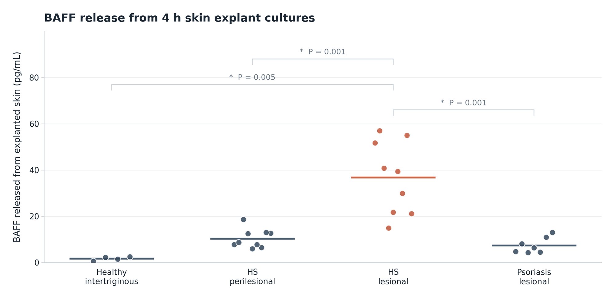
Beyond the directional comparison between the samples, concentrations in a medium say little without the volume of the medium and the size of the explanted sample. The supplementary materials state that the explants were 3 mm punch biopsies of whole thickness skin, but do not report the medium volume or how deep the punches reached. So these two parameters have to be estimated. HS biopsies for research should reach into the subcutaneous fat. A published whole tissue HS explant model keeps 4, 6 or 8 mm punches on 2 mL of medium per well. The medium volume was therefore swept from 0.25 to 2 mL and the depth of the 3 mm punches from 2 to 8 mm.
Across these ranges the lesions would release 40 to 760 pg of BAFF per mL of tissue per hour, with a median near 180 pg per mL and hour. Dividing the estimated quantities by four hours probably overstates the rate, because BAFF already present in the tissue washes out as well. Sabat et al. further cultured five lesional biopsies for 18 hours, in a medium that itself contained 2 percent human serum and therefore some BAFF of its own, and reported a BAFF release of 27 to 120 pg/mL with a mean of 54 pg/mL, while a constant release at the estimated release rate based on the four hour assay would have put the 18 hour value near 166 pg/mL. So my model probably overstates the actual release rate. However, this error is conservative, as it makes the lesions look like a larger source of BAFF than they probably are.
Soluble BAFF circulates as a trimer of three 17 kDa subunits. In humans, half of a subcutaneous IgG depot drains from the injection site within 7 to 12 hours, going by measured drainage rates of 0.09 to 0.16 percent per minute. Since BAFF is a protein roughly a third the size of IgG, it should leave faster. Nevertheless, for the sake of conservative assumptions, the escape half-life from the lesion was swept between 1 and 12 hours. Normal skin holds 40 to 45 percent interstitial fluid by weight, measured in rats. The model sweeps 25 to 50 percent, which errs toward higher concentrations.
BAFF from blood also equilibrates into the interstitial fluid of the lesion. This inflow was set at 30 to 60 percent of the roughly 0.8 ng/mL Sabat et al. measured in blood plasma of HS patients, at or below the 50 to 60 percent that IgG reaches in skin. This results in an estimate of lesional BAFF between 0.7 and 17 ng/mL, with a median near 2.8 ng/mL (Fig 2).
Sabat et al. also report the immune cell count of lesional biopsies, the share of neutrophils among these cells, and how much BAFF cultured blood neutrophils secrete. The neutrophil release rate estimated from these parameters is roughly 500 times lower than the rate from the explanted samples, so the explant rate, which the model uses, is the higher and therefore the more conservative of the two estimates.
I could not find a published serum concentration of tibulizumab in patients. The poster on the phase 1 in Sjögren's syndrome, dosed at 30 to 300 mg subcutaneously, shows observed and model-predicted profiles without giving exact values. The poster reports a half-life of 26.9 days, its abstract a subcutaneous bioavailability of 62.9 percent and a time to maximum concentration of 5 to 8 days.
So I built a one compartment model on these parameters. Half-life and bioavailability were drawn around them. The absorption rate was set to reproduce the published time to maximum concentration, and the volume of distribution was swept from 4.5 to 8 L, within the 3 to 8 L typical for antibodies. The half-life alone gives 1.9-fold accumulation on a four week interval.
In the phase 2 TibuSHIELD trial, 247 patients were randomized to 150 mg, 300 mg or placebo at weeks 0, 2 and 4 and then every four weeks. My model puts the steady state trough near 15 mg/L in plasma at 150 mg and near 30 mg/L at 300 mg. Antibodies reach skin at 15.7 percent of the plasma concentration by biodistribution coefficients and at 50 to 60 percent in interstitial fluid from mouse skin. The model sweeps 15 to 50 percent. At trough the lesion would then hold about 4.7 mg/L at the lower dose and 9.4 mg/L at the higher one, or 23 and 47 nmol/L for a 201 kDa molecule.
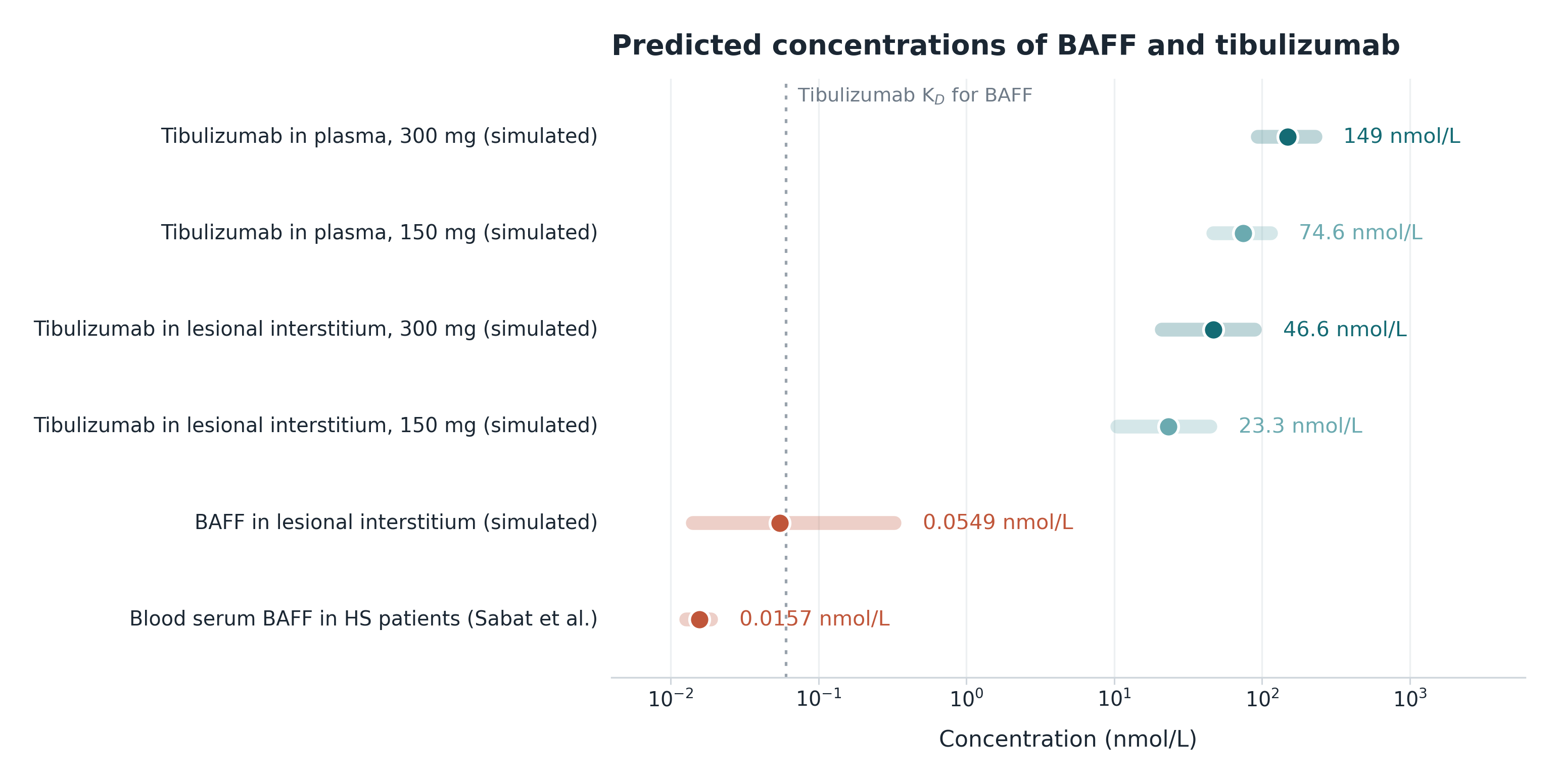
Lesional BAFF near 2.8 ng/mL is about 55 pmol/L of trimer. Tibulizumab would then be in roughly 400-fold molar excess over BAFF in the tissue at 150 mg and 800-fold at 300 mg. Across the draws the excess runs from 60 to 1,900 at the lower dose and from 120 to 3,900 at the higher one. The KD of 60 pM is the antibody concentration at which half of the BAFF is bound by tibulizumab at equilibrium. At the trough concentrations in the lesion, roughly 400 times the KD at 150 mg and 800 times at 300 mg, only about 0.3 percent and 0.1 percent of lesional BAFF would remain unbound. In other words, even at trough the antibody would bind more than 99 percent of the BAFF in the lesion.
Concentrations at one point in time are only part of the answer. Antibody-target complexes usually clear slowly, so drug is consumed at roughly the rate the target is produced. Tibulizumab has two anti-BAFF sites and two anti-IL-17A sites and binds both targets at the same time. A molecule holding BAFF should therefore still neutralize IL-17A (Caveat: the simultaneous binding was shown in vitro, I found no data showing this in patients).
TibuSHIELD requires at least five abscesses and inflammatory nodules. On ultrasound, the pseudocysts underlying many clinical nodules measure under 1 cm. A single wide excision removes 16.5 to 735 cm² of skin down to the fascia. All lesional tissue in one patient was estimated at 5 to 200 mL. Such lesions would then release 0.4 to 37 µg of BAFF over four weeks, which is 0.008 to 0.7 nmol of BAFF trimer.
At 62.9 percent bioavailability, a 150 mg dose of tibulizumab delivers roughly 470 nmol of antibody into the body, a 300 mg dose delivers 940 nmol. At one antibody per trimer, the median lesional production of 0.07 nmol would bind 0.02 percent of the lower dose and 0.008 percent of the higher one. The upper end of the range stays below 0.2 percent. Three antibodies per trimer would triple these numbers. Even a hundredfold higher lesional production than the explants suggest, from a higher release rate or a larger lesion volume, would bind about 2 percent of the lower dose of tibulizumab at the median.
After a single subcutaneous dose of the BAFF-binding drug atacicept, serum BAFF in healthy volunteers rose about 100-fold within two to four weeks to several hundred ng/mL, almost all of it bound to drug. Bound BAFF is cleared slowly, so the rise should show roughly how much BAFF the body produces in that time. Scaled to four weeks, a rise of 100 to 500 ng/mL in the 4 to 8 L of plasma and extracellular water in which antibody complexes are assumed to distribute corresponds across the draws to 1 to 5 mg of BAFF, or 20 to 100 nmol. Binding it would consume 2 to 10 percent of a 300 mg dose of tibulizumab per interval, or 4 to 20 percent of 150 mg. At the median, that is roughly 600 times the quantity of BAFF that HS lesions could release.
If these estimates are directionally right, the lesions add little to the BAFF in blood. Sabat et al. found blood BAFF in HS patients only slightly above that of healthy donors, without statistical significance, which is at least consistent with this reading. The 26.9 day half-life comes from the phase 1 trials, so systemic BAFF consumption was probably already part of the exposure the model is built on.
Like every simple model, this one has limitations and weaknesses, especially that it is mostly built on the results of a single ex vivo assay. The model treats lesions as uniform tissue. Abscess cavities and tunnels may hold material a punch biopsy culture misses. Membrane-bound BAFF should not reach the culture supernatant either, so the explant assay would not measure it. Quantitative proteomics counted 17,000 to 50,000 BAFF molecules per neutrophil in blood donors. That number covers the whole cellular content and probably overstates the surface pool. Even so, a lesional neutrophil count of a few hundred thousand per mL of tissue in 5 to 200 mL of lesions would add up to only about 0.001 nmol of BAFF per patient at the median. If every lesional neutrophil carried that whole pool on its surface, binding it would take about 0.0002 percent of a 150 mg dose, or 0.002 percent at the upper end of the draws.
Under the assumptions of this simple simulation, lesional BAFF is unlikely to consume a relevant share of tibulizumab. Hence the simulation suggests that tibulizumab could bind lesional BAFF by a comfortable margin. This simulation cannot predict whether tibulizumab will demonstrate efficacy in HS, but it suggests that the antibody is probably sufficiently dosed to bind lesional BAFF (which does not mean that targeting BAFF adds a relevant clinical effect in HS).
Follow me on X for frequent updates (@chaotropy).
Legal Information and Disclosures
General Disclaimer & No Financial Advice: The author is not a registered investment advisor or financial analyst, and nothing here is financial, legal, tax, or investment advice, a personal recommendation, an assessment of suitability for any investor, or an offer, solicitation, or recommendation regarding any security. This is not a securities research report and was not prepared under the requirements designed to promote the independence of investment research. It has not been peer-reviewed or audited, represents the author's opinion as of the publication date, and may change without notice. Readers should conduct their own due diligence and consult a certified financial professional before making any investment decision. Readers act on this content at their own risk, and to the fullest extent permitted by applicable law the author accepts no liability for any loss or damage arising from reliance on it.
Medical Disclaimer: The author is a physician, but this article discusses clinical trials and drug mechanisms as general scientific and educational commentary. It is not medical advice, diagnosis, or a treatment recommendation, and it establishes no physician-patient relationship. Readers should never disregard or delay professional medical advice because of anything written here, and should consult a qualified healthcare provider about any medical condition.
Computational Model: Every concentration this article reports for lesional BAFF and for tibulizumab in plasma and in lesions is the output of a simulation, not a measurement in patients, and Fig 2 shows simulated ranges rather than observed data apart from the blood BAFF row. Key inputs are not measured but swept across plausible ranges, as described in the text. The code has not been independently audited or peer-reviewed, and an error in it could change the results materially. Its outputs are order-of-magnitude estimates meant to show which quantities dominate, not predictions for any patient or trial.
Accuracy and Third-Party Data: The cited sources are believed reliable but are not guaranteed complete, current, or correct. The individual values in Fig 1, the 18 hour culture values, and the blood BAFF median and box in Fig 2 were read off the printed figures of Sabat et al. and approximate the published data points. The P values shown are those the paper reports. An exact Mann-Whitney U test on the digitized lesional and healthy values gives P = 0.003, the asymptotic form of the test gives the reported P = 0.005. That paper states that blood plasma was assayed while the axis of the corresponding panel is labeled serum. The explant culture volume, the depth of the punches and the cell density of the neutrophil cultures are not reported in the paper or its Online Repository and were swept or assumed. The tibulizumab pharmacokinetic parameters come from a conference poster and its abstract rather than from a peer-reviewed paper, none of the three completed phase 1 trials has posted results, and the dose levels of the phase 2 come from Zura's own slides because the trial record keeps them blinded. Despite the author's efforts to verify the sources cited, this article may contain errors of fact or interpretation.
Disclosure of Interest: The author holds a beneficial long position in Zura Bio Limited (NASDAQ: ZURA), and holds no position, long or short, in any other company mentioned. The author reserves the right to buy or sell securities of Zura Bio at any time without further notice. He received no compensation for this article, maintains no business relationship with any company mentioned, and has no access to non-public trial data, unblinded interim results, or internal company information. His position in Zura creates a potential for confirmation bias in the design of the model and in the interpretation of its results, and the choice of parameter ranges is where such bias would show first.
Forward-Looking Statements & Risk: Statements here about clinical results, regulatory outcomes, and market potential are predictions subject to significant risks and uncertainties, and actual results may differ materially. The TibuSHIELD topline has not been reported. A simulation suggesting that lesional BAFF is unlikely to limit drug exposure says nothing about whether tibulizumab will meet its primary endpoint, and the BAFF arm of the molecule has no controlled efficacy data in hidradenitis suppurativa. Readers should check Zura Bio's latest SEC filings. Biotechnology and pharmaceutical investing carries a high degree of risk, including total loss of principal, and past performance does not indicate future results.
Copyright: Original text and images are the property of the author and may not be copied, reproduced, or published in whole or in part without prior written consent, except as applicable law or the publishing platform permits. The figures are the author's own work. Fig 1 and the blood BAFF row of Fig 2 display values read from figures published by Sabat et al., and the underlying data remain the authors' and their publisher's. Use of this content to train machine learning or AI models requires explicit authorization. The title image is in the public domain and is not claimed as the author's property.